Osteonecrosis of the Hip
EspanolWhat is osteonecrosis of the hip?
Osteonecrosis or avascular necrosis is a disease where the blood flow to the head (ball of your hip joint, Figure 1) of the femur is interrupted that results in a painful hip.
Osteonecrosis literally means “bone death.” When the blood supply to the femoral head is damaged or disrupted, a portion of the bone dies. Pain and stiffness can occur. At first, only the ball of the femur, the femoral head, is involved. Over time, as the disease progresses, the joint surface of the femoral head may collapse (Figure 2) and lead to painful arthritis.
Symptoms of osteonecrosis typically present 12 to 18 months after injury to the bone or blood supply.
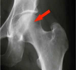 |
| Figure 1. Normal hip. Arrow points to the normal femoral head. |
What causes osteonecrosis?
Osteonecrosis can result from direct trauma to the hip, a systemic disease, or unrelated factors such as radiation. Sometimes the cause is unknown and is said to be idiopathic. When the disease results from injury, it is usually confined to the injured hip. When the disease is systemic, both hips and other joints such as the shoulders may be involved.
Trauma
Most of the blood supply to the femoral head comes through the lining of the hip joint called the capsule. This is a balloon like structure that inserts on the neck of the femur and keeps joint fluid in the hip. If the capsule is injured, the blood supply may be disrupted. When this happens there is no blood flow to a portion of the femoral head and the bone can die.
Injury to the capsule can happen in two ways. The first is traumatic dislocation of the hip where the ball of the femur is forced out of the socket (acetabulum). Usually, the dislocation is posterior, that is out of the back side of the hip. Posterior dislocations are commonly caused by high energy trauma such as an automobile accident or a fall from a height.
Printer Friendly
Click to print a copy of this article.
When there is a dislocation, it is important that the head be put back in the socket (reduced) as soon as possible. This can sometimes be done by closed manipulation under sedation or anesthesia. Other times, open surgery is required. Restoring the blood supply quickly can help prevent osteonecrosis
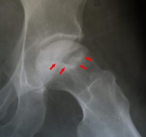 |
| Figure 2. Femoral head with osteonecrosis, outlined by arrows, notice the ball is no longer round. |
The capsule of the hip joint can also be injured by fracture. If the neck of the femur just below the femoral head is broken, and the fracture is out of place, it is likely that the capsule will be injured as well. When the capsule is damaged the fracture may not have enough blood supply to heal and osteonecrosis can develop. For this reason, displaced femoral neck fractures are often treated with hip replacement instead of fixing or pinning the break with screws. Even if a femoral neck fracture treated with pinning has healed, osteonecrosis can still be a secondary complication.
Systemic
Systemic causes of osteonecrosis are conditions which occur throughout the body. In this setting, it is common for both hips to be involved. It is not known if there is a single insult to the circulation or if the damage happens over a period of time. Some of the common systemic causes include:
Steroid therapy – It is not clear why taking steroids causes osteonecrosis. It is also not known what dose or length of steroid therapy causes harm. Many patients take steroids for conditions such as chronic lung disease, rheumatologic problems, colitis, or autoimmune diseases. If a patient has been taking steroids for a long period of time and develops hip pain, osteonecrosis should be ruled out.
Alcoholism – Although chronic alcoholism has been associated with osteonecrosis, the direct cause is unclear. It is thought that long-term alcohol use may damage some of the small vessels that supply the femoral head.
Autoimmune disorders – Diseases such as systemic lupus erythematosus, scleroderma, rheumatoid arthritis, and vasculitis are sometimes associated with osteonecrosis. As with steroid therapy and alcoholism, the reason for this is unknown. It may be due to the fact that many of these patients are given long-term steroids as part of their treatment.
Gaucher’s disease – Gaucher’s disease is a lipid storage disease where fatty tissues and diseased cells called Gaucher cells build up in organs such as the liver, the spleen, and bone marrow. The fatty infiltration may affect the blood supply to the hip.
Blood disorders – Diseases that affect blood flow or cause clotting disorders may cause osteonecrosis. Most common of these is sickle cell anemia Sickle Cell Anemia and Total Hip Replacement | Hip and Knee Care (aahks.org) where red blood cells become deformed into crescent shapes causing painful sickle cell crises in bone.
Other Causes
Caisson disease – Sometimes known as “the bends”, caisson disease can happen to divers who come to the surface too quickly after a deep dive. If they do not decompress slowly, nitrogen bubbles can form in small vessels and cause osteonecrosis. Typically, caisson disease is immediately painful after too rapid decompression.
Radiation injury – Patients who are treated with large doses of radiation may sometimes develop osteonecrosis in the radiated area as a result of their treatment. As with other factors the cause remains unclear but is most likely due to small vessel damage.
Transplantation – Osteonecrosis may occur in patients who have had organ transplantation. This may be related to anti-rejection drugs such as steroids.
What are the symptoms of osteonecrosis?
Pain is the earliest symptom of osteonecrosis of the hip. The pain is centered in the groin and radiates to the side and the back of the hip as well as the front of the thigh. It may radiate as far down as the knee. Sometimes it may feel like the pain is actually coming from the knee.
The pain may be present both with activity and at rest and typically will get worse when you are walking or bearing weight. Early on, you may have pain in your hip but still have good range of motion. It may feel like a pulled groin muscle that just doesn’t seem to go away.
In later stages, if arthritis develops in the joint, your hip may become stiff, and you may start to lose mobility. You may have difficulty putting on your socks or tying your shoes. It may be hard to get in a car and you may have to do stairs one at a time. The symptoms will be similar to those of osteoarthritis of the hip Osteoarthritis | Hip and Knee Care (aahks.org).
How do we diagnose osteonecrosis?
When you visit your doctor, he or she will take a history, ask you to describe your symptoms, and do a physical examination. Your doctor will then take x-rays which will most often make the diagnosis. Osteonecrosis of the hip has a characteristic appearance on x-ray. There is a wedge-shaped area of bone in the superior and lateral aspect of the head of the femur. The borders of the wedge are marked by dense bone (sclerosis) which has a whitish appearance on x-ray (Figure 2). The bone within the wedge will also appear abnormal.
On the lateral x-ray view, there may be a thin lucent (clear) line just beneath the surface of the joint. This is called a “crescent” or “meniscus” sign (Figure 3). In later stages of the disease, the joint surface may collapse.
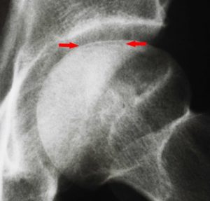 |
| Figure 3. X-ray of a hip with osteonecrosis with the arrows highlighting the clear line of the “crescent sign.” |
Early on, when a patient presents with only hip pain and the x-rays are normal, an MRI scan may be necessary to make the diagnosis (Figure 4). An MRI can tell how much of the bone is involved by the disease. It can also diagnose osteonecrosis in the opposite hip even though that hip may not have any symptoms at all.
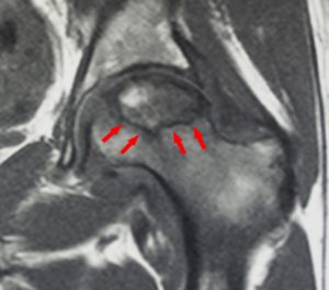 |
| Figure 4. MRI of a hip with osteonecrosis, arrows outline the wedge-like area of bone that is affected. |
How do we treat osteonecrosis?
The treatment of osteonecrosis depends upon the severity of your symptoms and how far the disease has progressed. In early stages, when pain is mild and does not interfere with function, anti-inflammatory medication and modified activity may be effective. However, once the diagnosis is made, it is important to assess the status of the femoral head. If the joint surface of the head is intact and has not collapsed, then surgical procedures that can restore and preserve the normal femoral head may be considered.
Surgical treatment of osteonecrosis of the hip
Often these procedures are done even though symptoms may not be severe. Early intervention may allow for new bone growth in the femoral head and prevent the need for hip replacement. The most common procedure is called a core decompression. A channel (or hole) is drilled from the base of the greater trochanter (point of your hip) on the side of the femur through the femoral neck into the area of osteonecrosis in the femoral head. The diseased bone is then reamed out and removed. This allows new bone to grow and replace the bone that has been removed. In some cases, bone graft material may be used to fill the area in an attempt to speed up the healing process. The graft may be an autograft taken from another area of the pelvis or an allograft, cadaver bone from a bone bank. Bone graft fills the space and creates a foundation for new bone growth in the femoral head.
Another option is to use a vascularized bone graft. This is a section of bone with an artery and vein attached. It is usually taken from the shaft of the fibula, the small bone of the leg. The artery and vein are then reattached to vessels around the hip. This provides a blood supply for the new bone.
Osteotomy is a procedure where the neck of the femur is cut, and the area of osteonecrosis is rotated away from the weight-bearing portion of the joint. This surgery is no longer performed very often because success rates are mixed, and the recovery is long. Once the joint surface of the femoral head has collapsed, core decompression and bone grafting will no longer be successful. When this happens, hip replacement is the treatment of choice.
When a total hip replacement is done, the diseased femoral head is removed, and replacement components (implants) are placed in both the femur and the hip joint socket. Implants | Hip and Knee Care (aahks.org).
In general, decompression and bone grafting procedures are best done in younger patients without large areas of osteonecrosis and without collapse of the femoral head. This is because it is best to preserve the natural hip, if possible. A replacement done at a young age may require a revision procedure later on. For older patients, the longevity of the prosthesis is less of an issue and replacement becomes a better option.
If you are being treated for osteonecrosis your orthopaedic surgeon can review the surgical options and discuss which procedure would be best for you.
What will my recovery be like?
The timing of recovery depends upon the surgical procedure. If you have had a core decompression or bone grafting procedure your surgeon may recommend that you use crutches and keep weight off your involved hip for up to three months after surgery. This is necessary to allow new bone to grow in your natural hip. When you begin bearing weight you may then need to go to physical therapy to help regain your strength and motion.
If a hip replacement is done recovery may be faster. You will be able to put weight on your hip immediately and progress from a walker or crutches to a cane in a shorter period of time. You will still need to do exercises or therapy to achieve a good result.
It is important to follow your doctor’s instructions in the recovery period to make sure you have the best possible outcome. You should also consider athletic restrictions and the need for long-term follow up with your surgeon.
This article has been written by Stuart Fischer, MD in collaboration with the AAHKS Patient and Public Relations Committee and peer reviewed by the AAHKS Evidence Based Medicine Committee.
Links to these pages or content used from the articles must be given proper citation to the American Association of Hip and Knee Surgeons.